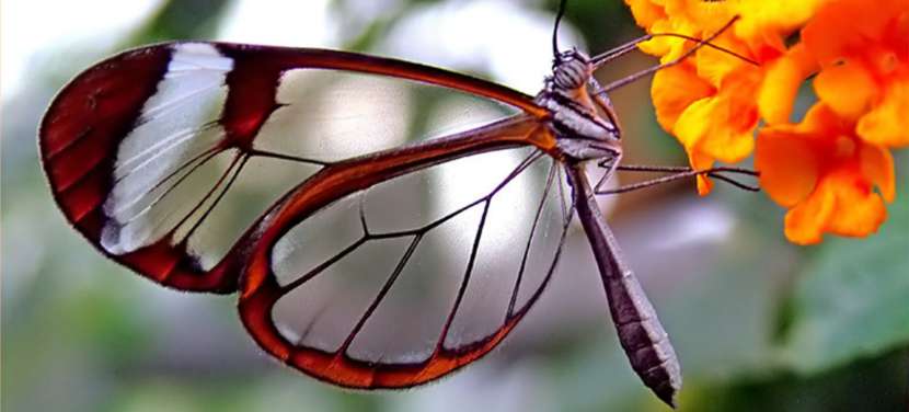
Perhaps this name does not sound familiar to you, or you know what it is because you have encountered such skin in your life or in your close surroundings. Epidermolysis bullosa is known as "butterfly skin" or "glass skin" and despite having a beautiful name, it is not a pleasant condition at all. This is a rare genetic disease that causes blisters and damage even with the slightest contact on the skin.
In individuals affected by the disease, the skin is extremely fragile, so a simple trauma, pressure from clothing, shoes, or even temperature changes can lead to blisters, painful sores, and erosions that require constant care. This extreme fragility resembles the wings of a butterfly, which is why it has received this name.
Beyond the visible effects on the skin, epidermolysis bullosa also brings complications such as mucosal issues, nutrition, growth, mobility, chronic pain, and infection risk; therefore, it requires careful clinical management and significant family and social support.
Butterfly skin is an inherited disease
Butterfly skin is a genetically based inherited disease, it is not contagious but currently has no cure. It does not spread through contact, sharing objects, or living together: it is not an infection, but a disruption in certain genes that connect the layers of the skin and provide its stability.
Additionally, it affects both the skin and mucous membranes such as the mouth, esophagus, perineal area, gastrointestinal system, respiratory system, and even the surface of the eyes. The sores caused by this disease appear like severe burns on the skin and repeatedly emerge with many complications.
Due to the persistence of open wounds, there is a high likelihood of infection, and individuals living with this disease may experience chronic nutritional deficiencies, anemia, and growth retardation. In some subtypes, the recurrence of sores and scars can also lead to loss of functionality in the hands and feet and an increased risk of certain types of skin cancer.
Children can also suffer from this disease because it is hereditary, being born with blisters or missing areas of skin. This is why it is called "butterfly skin," as the skin appears as delicate as a butterfly's wings. In newborns, suspicion often arises very early with the observation of blisters, erosions, or peeling of the skin after the slightest friction.
It lasts a lifetime
Children with "butterfly skin" will carry this disease throughout their lives, so they will need to learn to live with it from the early stages. This is not a temporary process; it is not an allergy that can disappear over the years.
There are different severity types of butterfly skin, and symptoms can vary; however, the basic clinical form does not change from mild to severe. This means that if a child has a mild variant, it will not turn into a very severe form, and if there is a severe form, it will not spontaneously turn into a mild one; however, good management can improve the quality of life.
The life and functional prognosis largely depend on the subtype of epidermolysis bullosa, the level of skin and mucosal involvement, infection control, nutritional support, and monitoring of long-term complications; for example, squamous cell carcinoma. In the mildest forms, many individuals reach adulthood with acceptable autonomy, while in more severe forms, there may be a high risk of morbidity and mortality from childhood.
Living with epidermolysis bullosa requires adapting daily life: making changes in clothing, shoes, how to hold babies, and the choice of activities and sports, as well as dedicating significant time for care, check-ups, and doctor visits. A psychological and social approach is critical for children and families to learn to integrate the disease into their daily routines and to reduce its emotional impact.
Symptoms of butterfly skin
Like any disease, epidermolysis bullosa or butterfly skin has characteristic symptoms experienced by individuals with this rare condition. Depending on its type and subtype, symptoms of epidermolysis bullosa may include:
- Alopecia (hair loss, caused by blisters on the scalp).
- Blisters around the eyes and nose may leave thin or scarred skin areas.
- Blisters around the mouth and throat can lead to feeding problems, difficulty swallowing, and pain while eating.
- Blisters on the skin after very minor injuries can occur even with temperature changes or minimal friction.
- Blisters present at birth or in the first days of life.
- Dental problems due to tissue fragility, leading to severe cavities and enamel erosion.
- Respiratory problems, cough, and hoarseness due to mucosal involvement.
- White swellings or pimples (milium cysts) in areas where blisters have healed.
- Nail loss or deformed nails (dystrophic nails), especially on the feet.
Individuals with epidermolysis bullosa typically show blisters in areas with the most friction, which may include the hands, feet, knees, elbows, or torso. These wounds can be bleeding, scab-forming, easily infected, and can cause severe itching. While scratching may provide temporary relief, it leads to the emergence of new wounds and the infection of existing ones.
After a blister heals, milium cysts, atrophic scars, or hypertrophic scars may appear; this can lead to the fusion of fingers and toes (syndactyly), joint contractures, aesthetic deformities, and significant functional loss in holding objects, walking, or performing daily activities. All of this can result in a loss of autonomy if proper prevention and treatment are not undertaken.
In addition to skin symptoms, epidermolysis bullosa can trigger extracutaneous symptoms; this can affect skin appendages (nails, hair, teeth) and the gastrointestinal, urinary system, or lung epithelium. This leads to conditions such as feeding problems, constipation, recurrent urinary tract infections, respiratory difficulties, and increased fatigue.
Because the images of this disease can be quite harsh and striking in many cases, they are often avoided being shown without context. If someone wishes to see the effects of this disease, they can search on Google; it should be remembered that behind every image, there is a person and a family living with a complex disease.
Types of Epidermolysis Bullosa and Genetic Inheritance
Epidermolysis bullosa is a genetic disease that passes from parents to children. There are different forms of epidermolysis bullosa depending on the depth of the skin where blisters occur and the type of inheritance received by the child. In terms of inheritance, essentially two main models are distinguished:
- Dominant inheritance. This type of inheritance occurs when one of the parents also carries the disease and directly transmits it to their children. In this case, the probability of the child inheriting the disease in each pregnancy is approximately 50%, as a single defective gene copy is sufficient for the disease to manifest.
- Recessive inheritance. In this case, the parents are healthy carriers of the gene that causes the disease, but they do not experience the disease themselves. They are the ones who pass the disease to their children, and the likelihood of the child having epidermolysis bullosa is 1 in 4 pregnancies; there is a 50% chance of being a healthy carrier and a 25% chance of not inheriting the disorder. Affected children and carrier children can only have affected children if their partners also carry the epidermolysis bullosa gene. In this context, there is a 25% chance of having a healthy child, a 50% chance of being a carrier, and a 25% chance of having an affected child in each pregnancy.
At the molecular level, many forms of epidermolysis bullosa are linked to mutations in genes that code for proteins vital for the connection between the epidermis and dermis. A prominent example is the COL7A1 gene, which contains instructions for synthesizing type VII collagen, a critical protein for skin cohesion. Hundreds of mutations associated with the disease have been identified with this gene; in both dominant and recessive forms.
Clinically, accurate genetic classification allows for predicting the severity of the disease, the risk of skin cancer, long-term evolution, and treatment options, including research involving some advanced treatment methods. Therefore, genetic counseling is of fundamental importance for families to understand the risk of recurrence in future pregnancies.
Subtypes of butterfly skin
Approximately twenty subtypes have been identified for epidermolysis bullosa or butterfly skin, each with its own distinctive clinical features. Different forms can be grouped into three main types considered most significant; however, mixed forms are also described in practice:
- Simplex. This type of epidermolysis bullosa is characterized by tearing in the top layer of the epidermis (intraepidermal level). Blisters heal without significant tissue loss and usually heal without leaving deep scars. Affected individuals may experience a certain sense of healing over time, but the disease never fully resolves. It is generally the most common form and is mostly limited to the hands and feet, particularly in cases of friction or temperature.
- Junctional. This type of epidermolysis bullosa occurs when blisters arise at the dermoepidermal junction, in the lamine lucidum layer of the basement membrane. Subtypes vary from very severe forms in the neonatal period to others that improve somewhat with age, but do not disappear. It is a rare form, but is associated with extensive involvement of the skin and mucous membranes, and typically requires specialized care and multidisciplinary follow-up.
- Dystrophic. In this type, blisters form in the deepest layer of the skin, beneath the basement membrane, in the dermis. As they heal, wounds can lead to joint restrictions, fusion of fingers (syndactyly), and significant limitations in mobility. Additionally, blisters can form in the pharynx, mouth, esophagus, stomach, intestines, respiratory and urinary tracts, and even in the eyelids and cornea. This form is associated with an increased risk of squamous cell carcinoma at an early age.
In addition to these, a rare form of epidermolysis bullosa known as Kindler syndrome has been described, characterized by fragility of the skin, pronounced photosensitivity, progressive pigment changes, skin atrophy, and thickening of the palms and soles. In this syndrome, the separation of skin layers can occur at different levels (multilevel), distinguishing it from other classical types.
How does epidermolysis bullosa affect the skin?
Depending on the exact location of the blister formation in the skin, the disease is divided into several major groups and also into different defined subgroups. The details at this level are not merely theoretical: they have practical implications and can affect the prognosis, the type of care required, the frequency of complications, and the risk of permanent effects.
In simple forms, when tearing occurs within the epidermis, the skin tends to regenerate with less scarring, but there may be pain, itching, and significant discomfort. In junctional forms, the lesions are located in the basement membrane and present with extreme fragility from birth, involvement of mucous membranes, and early intensive care needs.
In dystrophic variants, separation occurs beneath the basal membrane, at the subepidermal level. Therefore, the blisters leave hard and attractive marks and over time begin to pull on the surrounding skin. This leads to restrictions, abnormal joint flexions, syndactyly, and increasingly limited ability to use hands, walk, or perform simple tasks.
In mixed forms, the same patient may exhibit characteristics of multiple levels of effect, which complicates diagnosis and management. The risk of aggressive skin cancer is highlighted in chronic wounds; therefore, the importance of regular dermatological follow-up is emphasized.
Complications of butterfly skin
Butterfly skin can also lead to many local and systemic complications, such as:
- Infections, due to blisters and open wounds, which can be bacterial, viral, or fungal.
- Sepsis. Sepsis occurs when bacteria from an infection enter the bloodstream and spread throughout the body; this threatens the person's life and requires emergency hospitalization.
- Deformities. Conditions such as fusion of the fingers of the hands or feet (syndactyly), abnormal flexions in the joints, skin pull, and reduced mobility.
- Malnutrition and anemia. Blisters in the mouth, tongue, or esophagus can make eating and drinking very difficult; this leads to low food intake, weight loss, vitamin and mineral deficiencies, anemia, and delays in the healing process. In children, slowing growth rates and developmental disorders may also be observed.
- Dehydration. Open and large blisters can cause significant loss of body fluids, leading to severe dehydration, especially in infants and young children.
- Constipation. The presence of painful blisters in the anal or perineal area may cause some patients to postpone bowel movements; this leads to severe constipation. It may also be due to insufficient fluid, fiber, fruit, and vegetable intake, which can stem from fear of pain or difficulty swallowing.
- Eye problems. Chronic inflammation of the eyes and eyelids can damage the cornea, cause recurrent erosions, and sometimes lead to vision loss.
- Skin cancer. In certain dystrophic types, adolescents and adults can develop a cancer called squamous cell carcinoma on chronic lesions or non-healing wounds.
- Atrophy and changes in skin color. In some subtypes, the skin becomes increasingly thinner, with areas of hyperpigmentation and hypopigmentation starting on the upper hands and neck, spreading to other parts of the body.
- Thickening of palms and soles, leading to the formation of painful calluses that make walking and using hands difficult.
- Loss of skin lines, including fingerprints, which can complicate identification in biometric systems.
- Other gum involvement, inflammation of the gums, tooth loss, and excessive growth of surrounding gums.
- Intestinal inflammation in some subtypes worsens digestive and nutritional problems.
- Death. Infants with severe forms of epidermolysis bullosa are at risk of serious infections, malnutrition, and significant fluid loss. Difficulty eating and breathing can severely impact survival. Unfortunately, many of these children die in childhood despite all available care.
It is very important to follow the doctor's instructions and adhere to the prescribed treatment according to the condition of epidermolysis bullosa in daily life. This includes elements such as wound care, pain control, nutritional support, physical therapy, eye monitoring, and oncological follow-up.
Diagnosis of epidermolysis bullosa
A healthcare professional may suspect the presence of epidermolysis bullosa based on the characteristic appearance of the skin and medical history: blisters after the slightest friction, wounds from birth, family history, or the presence of abnormal lesions.
To confirm the diagnosis and determine the specific subtype, several laboratory tests are usually required:
- Immunofluorescence mapping with skin biopsy allows for the identification of the separation site between the epidermis and dermis and the assessment of the presence of specific adhesion proteins.
- Electron microscopy in selected cases is used to view the structure of the basement membrane and adhesion fibrils in detail.
- Genetic molecular tests are necessary today to identify the responsible mutation, classify the subtype, predict prognosis, and provide appropriate genetic counseling to the family.
Genetic diagnosis helps to early identify the severity of the disease, make decisions about patient management, inform the family about risks in future pregnancies, and assess the likelihood of access to targeted therapies or clinical trials based on specific genetic changes.
Common treatment and daily care
Today, despite being extensively researched, epidermolysis bullosa does not have a definitive treatment. Treatment focuses on preventive and symptomatic measures for skin lesions and systemic complications.
When the disease manifests, rapid and coordinated intervention is vital; as it significantly affects the quality of life and lifespan of patients. The approach is often multidisciplinary, involving dermatology, pediatrics, wound care nursing, nutrition, physical therapy, dentistry, ophthalmology, psychology, and sometimes plastic surgery and traumatology.
The cornerstones of treatment are as follows:
- Wound care, with non-adherent dressings, careful techniques to drain some blisters, measures to reduce friction, and prevention of infections.
- Pain management, using analgesics tailored to age and severity of symptoms, both during care and in daily life.
- Nutritional support, high-calorie and protein diets adapted to chewing or swallowing difficulties, supplements as needed, and feeding tubes or other supportive measures in severe cases.
- Physical therapy and rehabilitation, to prevent and treat restrictions, maintain joint mobility, and delay the onset of deformities.
- Dental and oral care, regular check-ups, special hygiene measures adapted to the fragility of the gums, and restorative or preventive treatments to reduce cavities if necessary.
- Psychological and social support, for both the affected individual and their family, is important considering the emotional impact and burden of daily care.
New research areas and advanced treatment methods
In recent years, significant advances have been made in the field of genetics and genomics regarding epidermolysis bullosa. New treatment strategies are being investigated, including gene therapy, cell therapy, transplantation of genetically modified epithelial cells, and tissue engineering to modify damaged skin areas.
Some of these treatments have shown very promising results, especially in certain recessive dystrophic subtypes. The most active research areas are:
- Topical gene therapies, in the form of gels or lotions that integrate healthy genes into skin cells, to partially restore the function of adhesion proteins.
- Transplantation of epithelial cells modified by gene therapy, applied as skin grafts in severely affected areas.
- The use of viral and non-viral vectors to place functional gene copies into the patient's cells, for genes such as COL7A1.
Although these options are not yet generally available, ongoing research is opening the horizon for more specific and personalized treatments in the future. In the meantime, current management is primarily based on daily care and prevention of complications.
The impact on daily life and support for families
Butterfly skin not only requires medical knowledge but also necessitates holding babies, choosing clothes, organizing care, preparing nutrition, and planning education as part of a lifestyle change. Activities that are simple for other families can turn into a daily struggle in situations where every friction can cause a new wound.
Care procedures can take several hours a day and require special materials, time, patience, and pain management. Simple movements like walking, writing, playing, or eating can be painful at certain moments; therefore, the environment must adapt and show understanding.
The support of patient associations, AEBE DEBRA (Spanish Epidermolysis Bullosa Association), is particularly valuable. These organizations provide up-to-date information, resources, emotional support, social counseling, and opportunities to share experiences with other families facing similar situations.
You can find more information about butterfly skin through AEBE DEBRA (Spanish Epidermolysis Bullosa Association); here, the types of the disease, available resources, and the latest developments in research are explained in detail.
Butterfly skin or epidermolysis bullosa is a rare but profoundly complex disease that affects much more than just the surface of the skin. Knowing its genetic causes, different clinical forms, associated complications, and treatment possibilities allows for safer care, alleviating daily pain, and better accompanying those living with such delicate skin.


Comments
(4 Comments)