Perhaps a friend has told you that they had to give birth by cesarean section because their baby experienced umbilical cord prolapse in the final moments of delivery.
Although it is not very common, as healthcare professionals working in deliveries, we are quite familiar with this situation. Understanding what this complication is helps to navigate the pregnancy and delivery process more peacefully and ensures that you are informed about when to seek help in emergencies.
To understand umbilical cord prolapse, it is important to know what the umbilical cord and its functions are, how it behaves within the amniotic sac, and what changes when labor begins.
Umbilical Cord
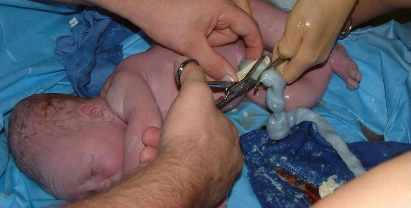
The umbilical cord is the structure that connects the baby to the placenta. Working like a true "lifeline," it facilitates the continuous exchange of oxygen, nutrients, and waste products.
It forms from approximately the 5th week to the 12th week of pregnancy and plays a fundamental role in the baby's development. It begins to function even in the very early stages of pregnancy, ensuring that the embryo and later the fetus receive everything they need to grow.
The umbilical cord is a flexible and durable tube. Inside it, two arteries and one vein are coiled in a spiral shape. All of this is protected by a jelly-like substance called Wharton's jelly; this substance helps support, bind, and protect the three vessels, preventing them from collapsing easily.
The length of the cord varies, but it is generally around 50 centimeters and weighs about 100 grams. In some cases, it may be shorter or longer, and most of the time it does not cause problems, but an abnormally long cord increases the risk of looping, knotting, or prolapse.
Normally, the cord attaches to the placenta in the center, but it can also attach more laterally. Atypical placements, such as a velamentous insertion where vessels reach the placenta through membranes, can be associated with other risks, but are generally not the direct cause of cord prolapse.
Functions of the Umbilical Cord
The umbilical cord is the connection between the baby and its mother. It connects to the placenta through the cord; the placenta takes oxygen, glucose, and other essential nutrients from the mother's blood and sends back the waste products produced by the baby.
Interestingly, the arteries of the umbilical cord carry venous blood; that is, they carry blood filled with carbon dioxide and waste products from the baby's body to be purified at the placenta. The umbilical vein carries oxygenated blood necessary for the baby's growth.
Additionally, the cord serves a mechanical function: thanks to its length, flexibility, and Wharton's jelly, it allows the baby to move within the womb without cutting off blood flow; this explains why most loops or twists of the cord do not create clinical issues.
What Happens During Pregnancy?
During pregnancy, the umbilical cord grows longer with the growth of the baby and the placenta. Its final length is related to the freedom of movement of the fetus: babies that are more active and have more space may have slightly longer cords.
The umbilical cord is a relatively long channel that does not require the baby to remain attached to the placenta for feeding or waste disposal. The cord allows these functions to be performed at a certain distance; this enables the baby to gain freedom of movement within the womb and facilitates proper development.
Thanks to the protection of amniotic fluid and Wharton's jelly, the baby can turn, stretch, or even play with the cord; in most cases, blood flow is not compromised.
During pregnancy, the placement of the cord can sometimes be seen with ultrasound, but this alone does not predict whether there will be complications during delivery. The cord wrapping around the neck (nuchal cord) or around a limb does not necessarily mean a serious risk during pregnancy.
What Happens in the Amniotic Sac?
The umbilical cord and the baby float in amniotic fluid; this is a "natural pool" that protects the fetus, facilitates its movements, and prevents the cord from being subjected to significant compression. This floating allows the cord to withstand fetal movements well during pregnancy, thanks to the effect of Wharton's jelly.
When the protection ends, especially in the case of rupture of the amniotic sac and a sudden decrease in fluid volume, problems may arise. In this case, the cord may slip down towards the lower part of the uterus and can enter the birth canal if the fetal presentation is not well positioned.
Therefore, the position of the baby and the placement of the initial presentation (head, buttocks, shoulder, etc.) affect the likelihood of cord prolapse during delivery.
What is Umbilical Cord Prolapse?
During delivery, the umbilical cord should not be compressed by any structure. Compression can cut off blood flow between the placenta and the baby, leading to oxygen deprivation.
Ideally, the cord should always be above the baby's head. Umbilical cord prolapse occurs when the cord remains in front of the fetal presentation (usually the head); that is, it can slip between the baby's head and the mother's pelvic bones or even through the cervix into the vagina.
For this situation to occur, the amniotic sac usually needs to be ruptured; this can happen either spontaneously or artificially. If the fluid suddenly escapes, it can pull the cord down and drag it downward before the baby's head is well positioned. It can also occur later if the position of the head changes during delivery and creates a space that causes the cord to slip.
Types of Umbilical Cord Prolapse
Experts generally distinguish two main forms:
- Open or obvious prolapse: the cord falls in front of the presenting fetal part and is felt or seen during a vaginal examination or protruding from the vulva. It usually occurs after the membranes rupture and is an obstetric emergency.
- Hidden prolapse: the cord is trapped between the baby and a structure of the mother (usually the pelvis), but it is not visible. It is usually suspected with changes in fetal heart rate; this includes conditions like bradycardia or prolonged variable decelerations.
In both cases, the underlying issue is compression of the cord, which prevents adequate blood flow to the baby.
Is Umbilical Cord Prolapse Serious?
If the fetal part is compressing the cord, the flow of oxygen and nutrients may be partially or completely cut off; this can lead to fetal hypoxia. Prolonged compression can cause acute fetal distress, acidosis, and if not acted upon quickly, can lead to neurological damage or death.
Therefore, cord prolapse is considered an obstetric emergency that requires immediate intervention. Fortunately, this condition is rare, and with prompt diagnosis and protocols, most babies are born without significant lasting damage.
What Are the Causes and Risk Factors of Umbilical Cord Prolapse?
Cord prolapse is more likely to occur when there is more space in the pelvis or an abnormal relationship between the baby and the birth canal. Risk factors include:
- Premature birth: the head may not be well positioned in the case of rupture of the sac.
- Low birth weight: a smaller baby leaves more room for the cord to slip down.
- Multiple births.
- Abnormal presentations (breech, transverse, etc.).
- Premature rupture of the amniotic sac, especially if the presentation is not well positioned.
- Polyhydramnios (excess amniotic fluid) facilitates the sudden exit of the fluid.
- Abnormally long umbilical cord.
- Placental abnormalities or unusual placements of the cord.
- Having multiple births or a wide maternal pelvis.
- Obstetric maneuvers (external version, artificial rupture of membranes without proper monitoring, etc.).
Having one or more risk factors does not mean that prolapse will occur; however, it requires careful attention, especially during membrane rupture.
What Are the Symptoms of Umbilical Cord Prolapse and How Is It Detected?
In many cases, prolapse is diagnosed in the hospital during delivery. Sometimes the woman feels nothing, and the medical team detects it through examination or monitoring.
Symptoms and diagnostic methods:
- Visible or palpable descent of the cord: can be seen during a vaginal examination or may protrude from the vulva after membrane rupture.
- Changes in fetal heart rate: variable decelerations, bradycardia, or abnormal fetal monitoring on the monitor (CTG).
- Decrease in fetal movements: a late sign of fetal distress, but always requires urgent attention.
If you are at home and feel that the sac has ruptured and something is coming out of the vagina, or if you feel a strange sensation or sudden fluid loss, go to the hospital immediately and report the situation you observed. Quick response is very important.
Possible Complications and Outcomes
If not detected or treated quickly, cord compression can lead to:
- Fetal hypoxia and irreversible brain damage within minutes in severe cases.
- Acute fetal distress and the need for emergency intervention.
- Neurological complications in the newborn (cerebral palsy, developmental disorders) following severe perinatal asphyxia.
- Intrauterine or perinatal fetal death, in the most extreme cases if intervention is not performed quickly.
The most significant outcome for the mother is usually the need for emergency cesarean section; this carries risks such as bleeding, infection, and a longer recovery time, but the priority is always to protect the life and health of the baby.
Medical Intervention and Treatment in Case of Umbilical Cord Prolapse
Cord prolapse usually occurs during delivery in the hospital. Speed of intervention is crucial as it is necessary to prevent the continuation of compression and its negative effects on the baby.
Common measures:
- Immediately changing the mother's position: positions supported by the knees and torso (knee-chest) or the Trendelenburg position can help move the head away from the birth canal and reduce cord compression.
- Manually elevating the presenting fetal part: the specialist can insert their hand to hold the head slightly elevated and reduce pressure on the cord while preparing for cesarean.
- Providing oxygen to the mother and other supportive measures aimed at temporarily improving fetal oxygenation.
- Continuous fetal monitoring; this is necessary to assess the response to maneuvers and determine the urgency of delivery.
- Emergency cesarean: when the cord is clearly in front of the head or if there is fetal distress, it is usually necessary to prevent harm.
If the head is well positioned and the cord is only on the side, it may sometimes be possible to gently place the cord and continue with careful monitoring. However, if there is any doubt about fetal well-being, the safest option is immediate cesarean.
The quick coordination of the obstetric team and the presence of clear protocols in the delivery rooms make a significant difference in prognosis.
Knowing what the umbilical cord is, how it works, and why umbilical cord prolapse is an emergency helps to better understand the decisions that the medical team can make during delivery. Although it is a rare complication, being well-informed and quickly going to the hospital in case of membrane rupture or if you feel something unusual in the vagina are important steps to ensure that your baby is born in the best conditions with the help of professionals.


Comments
(7 Comments)